Guiding brain surgery with evoked electrophysiology

The goal is to use direct electrical stimulation (DES) evoked electrophysiology of the brain during brain surgery to diagnose and determine the location of the tumor or epileptogenic zone and to better understand online anatomical connectivity to guide surgery in awake patients or those under general anesthesia
Underway
01/01/2021

-

31/12/2025

Main objectives

By combining experimental measurements and modeling our goal is to:
- Develop the recording of evoked potentials and optimize SED to make the method reliable and reproducible, and allow real-time and in vivo measurement of local electrophysiological state (e.g. excitability) and connectivity. In other words, to develop electrophysiology imaging of healthy and diseased tissues.
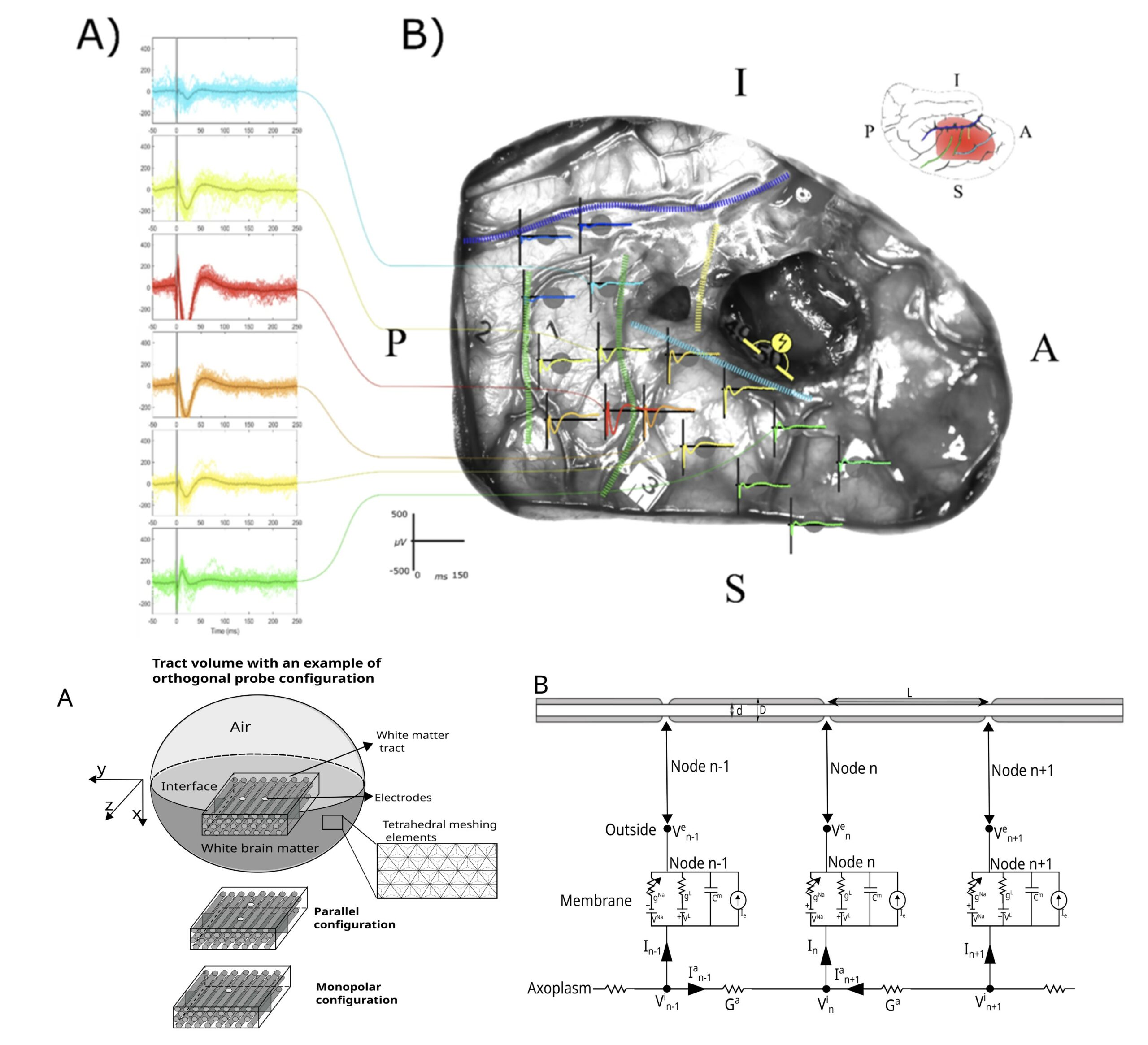
- While modeling the effects of electrical stimulation and its diffusion, mainly from axon models
Top: Experimental measurements and evoked potentials measured by Electrocorticography (ECoG) during SED applied to the white matter bundles. Bottom left: Partitioned 3D conductive model. The current injected in the 3D model at the level of the electrode contacts imposes an electric potential field. The definition of this potential field is done by a finite element method: a tetrahedral mesh is created where on each vertex of the tetrahedron the electric potential is calculated in quasi-static condition. Bottom right: Electrical circuit of the dynamic membrane model and the cable equation. The central node gives the description of the currents involved in the resolution of the membrane potential. The bottom line describes the contribution of axoplasmic currents to this central node

Challenge

Direct electrical stimulation of the brain (DES) has been used for almost a century as a tool for intraoperative functional exploration in an awake patient (Penfield 1937, 1947). The principle is as follows: by transiently applying a focal electrical current, the patient's functioning is transiently disturbed. This tool allows real-time mapping of essential functional regions (language, motor skills) and is now successfully used in clinical practice in awake surgery of epileptogenic foci and brain tumors: the resection can be pushed safely to the functional limits. Despite its proven clinical efficacy at the behavioral level, very little is known about the electrophysiological effects of electrical stimulation. This understanding is also crucial for the emerging field of new technological neurodevices used in acute (awake surgery) and chronic (neuromodulation, neuroprostheses and brain-machine interfaces)

Methodology/Technology used

Method of recording and processing evoked potentials by electrical stimulation applied to the brain. Biophysics of electrical stimulation, axon models and cable equations

Publications

Contacts

Emmanuel Mandonnet () & François Bonnetblanc ()

Members

AP-HP, Inria, Hôpital Gui de Chauliac (Montpellier), Kobe University Graduate School of Medicine, Japan

Useful links

https://project.inria.fr/inriaassociateteamjapanfrance/team/

Other projects

COVIPREDS

Description

US Caractérisation et prédiction de la survenue de formes graves ou létales du COVID-19 à partir des données issues de l’EDS de l’AP-HP

Names of partners involved
AP-HP, Inria & Centrale Supélec

read more